Research Supported by CGF
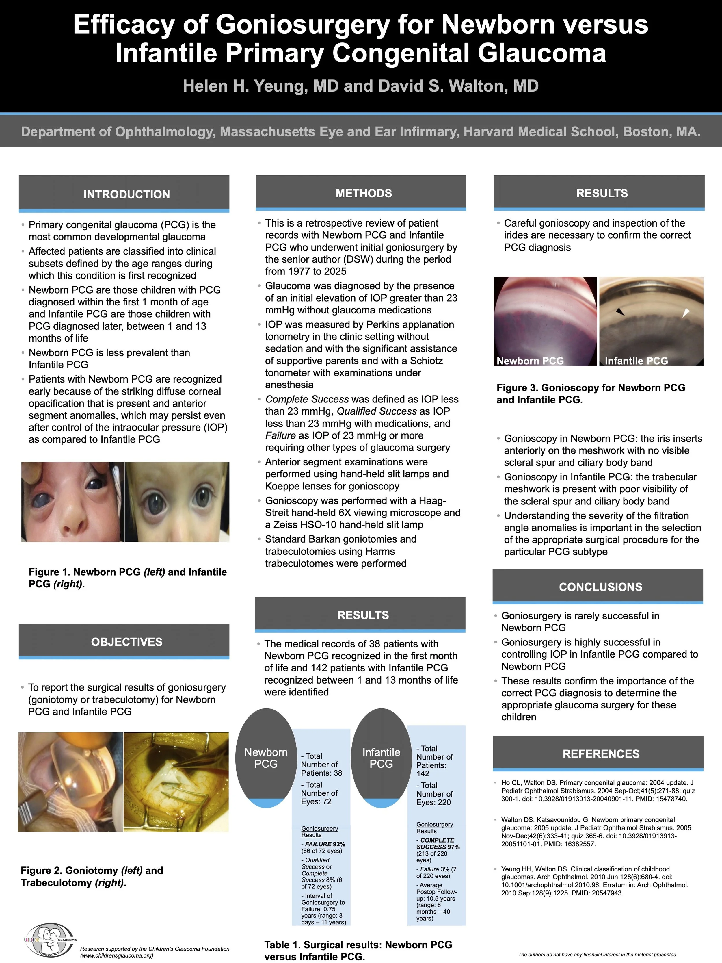
Poster Presentation at the American Association of Pediatric Ophthalmology and Strabismus 2026 Annual Meeting
Boston, MA on April 2026
Publications —
Infantile Aphakic Glaucoma: A Proposed Mechanism
Helen H. Yeung, MD; Rajendra Kumar-Singh, PhD; David S. Walton, MD
Click here to View Publication
Ongoing Research —
University of Georgia
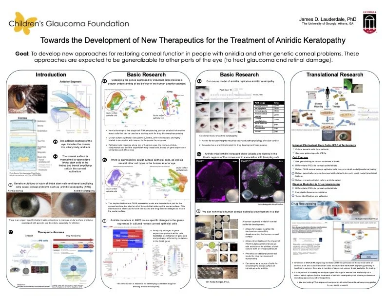
Preventing Vision Loss in Aniridia by Targeting Early Corneal Instability
Aniridia-related keratopathy (ARK) is a progressive eye disease that typically leads to corneal scarring and loss of transparency, resulting in vision loss. It occurs in individuals with reduced functional levels of the gene PAX6, which is essential for maintaining corneal health. Currently, no therapies exist that prevent disease onset or progression. Most individuals with aniridia (approximately 80–90%) develop ARK, although the timing and severity vary widely between patients and even between eyes. This variability suggests that disease progression is not fixed but instead depends on additional biological triggers.
ARK is generally classified as a progressive limbal stem cell deficiency leading to conjunctivalization and corneal opacity. However, our data suggest that early disease is not solely driven by stem cell deficiency, but instead reflects a stable epithelial state characterized by identity instability, barrier dysfunction, and low-level immune priming that remains compatible with corneal transparency. Our RNA sequencing data confirm that these corneas exhibit early stress and “repair-like” gene expression without activation of the fibrotic and inflammatory programs that drive opacity.
We hypothesize that ARK develops in two stages. In the first stage, reduced PAX6 destabilizes corneal epithelial identity but remains compatible with transparency. In the second stage, inflammatory signaling triggers a transition to fibrosis, vascularization, and tissue remodeling, resulting in vision loss.
To test this hypothesis, we will use RNA sequencing and targeted analyses to define molecular signatures that distinguish stable from progressive corneal states. We will then test two complementary therapeutic strategies in a mouse model: (1) enhancing PAX6-dependent epithelial stability pathways to restore corneal homeostasis, and (2) inhibiting key disease-driving signaling pathways, including MEK/ERK-mediated inflammatory amplification and CTGF-driven fibrosis. We will measure outcomes at both the molecular and tissue levels, linking gene expression changes to corneal clarity, structural remodeling, and disease progression. A central goal is to determine whether early molecular stabilization can delay or prevent the transition to opacity. This work shifts the focus from treating end-stage damage to intervening during a previously unrecognized, pre-disease state. By identifying the molecular events that control when ARK progresses, we aim to develop strategies that extend corneal clarity and preserve vision.
In summary, this project reframes ARK as a condition in which disease is common but temporally variable and where targeting the transition from stability to pathology offers a critical opportunity for prevention.
-Dr. James D Lauderdale
Associate Professor
The University of Georgia
Department of Cellular Biology
Past Research —
Tufts University School of Medicine
Aphakic Glaucoma Research
“In last year’s newsletter of The Children’s Glaucoma Foundation (2019), you read about a new research initiative to develop a gene therapy approach for the treatment of infantile aphakic glaucoma. I am excited to report that since the initiation of those studies, we have made tremendous progress. Specifically, we have completed development and testing of a novel adeno-associated virus (AAV) that expresses an inhibitor of a process known as epithelial to mesenchymal transition (EMT) of lens epithelial cells. AAV is currently an FDA-approved gene delivery method for a disorder in children that causes blindness and hence we believe that while the path we are taking is highly novel, it is de risked by prior success in this field.
The process of EMT leads to a blockage of the outflow pathway of fluids in the front of the eye due to ‘clogging’ of tissues known as the trabecular meshwork. This blockage to outflow results in a build-up of pressure in the eye that ultimately results in the death of cells in the back of the eye and consequently, blindness. The FDA requires that novel therapies be tested for safety and efficacy prior to testing in humans. During the previous year, we have modified the local environment of the eyes of small rodents such as to increase the pressure within the eye. This surprisingly led to very high levels of EMT at the trabecular meshwork and a significant increase in pressure within the eye. We essentially generated a non human model of infantile aphakic glaucoma. We also tested this same disease process in the presence of our therapeutic AAV and we found that both the EMT and pressure within the eye was reduced. These results are extremely exciting. Before proceeding, we need to test various doses of the therapy and in enough examples such that they are highly statistically significant. We had anticipated completion of these studies by Summer of 2020 and although we are still on target, the temporary shutdown of our laboratories due to Covid-19 has set us back by about three months. As of writing, I am pleased to report that the studies have been re initiated and we are moving forward. Additional excitement comes from having tested and proven our AAV vector in a larger eye akin to that of a human. We essentially already have proof-of-concept of our hypothesis. During the next 4 to 6 months we expect to complete these studies and start planning the generation of GMP (good manufacturing practice) grade AAV vector needed for obtaining safety data that would be used for applying to the FDA to test a new investigational drug (IND) in humans. These latter studies will collectively take two to three years but our unprecedented success thus far gives us hope that we can complete them sooner rather than later.”
-Rajendra Kumar-Singh, PhD
Professor, Developmental, Molecular and Chemical Biology
Tufts University School of Medicine